
Diagnostics & Therapy
The right treatment for every symptom.
For a healthy and relaxed chewing device.
In optimal condition, the upper and lower rows of teeth meet evenly. Chewing muscles, joints and teeth are then in harmony. If this system is disturbed, the entire body statics can get out of balance.
A strong back bite leads to head retention and changes the position of the cervical spine.
This can block the pelvic area and lead to a difference in leg length.
Impairments such as shoulder-arm syndrome, headache, tinnitus, sciatica or knee problems are just as related to TMD as night-time teeth grinding due to psychological stress.


We will be glad to help!
The complaints or clinical symptoms of TMD, i.e. a disorder between the articular processes of the lower jaw and the corresponding articular surfaces on the head, can occur on the spot (close to the joint) and thus clearly identifiable, but also at other parts of the body (away from the joint).
The joint-related complaints, such as pain, cracking and restricted movement of the temporomandibular joints, are usually quickly recognized by dentists. The cause can be changes in tooth position due to tooth loss or restorative measures through crowns or dentures.
The complaints of the TMD that are far from the joints are noticeable through chronic headache, neck pain or facial pain as well as tinnitus, but also through non-specific vegetative disorders. For many medical professionals in various specialties, these symptoms are often not recognizable as the consequences of TMD.
Therefore, neurophysiological diagnostics play an important role in therapy preparation and follow-up check-ups.

Diagnosis
There are many ways to reach the desired results
In our CMD-Center, experts from many different disciplines work hand in hand. We have a variety of examination options for our patients and their diagnosis of CMD.
For example the manual function analysis, which is carried out without the aid of technical tools, and the opposite instrumental function analysis.
Other diagnostic methods would be MRI (magnetic resonance imaging) for imaging soft tissue structures and digital volume tomography (DTV).
Manual functional Analysis
The manual functional analysis is conducted without aid of technical devices or instruments. Putting manual stress on the muscles and joints of the masticatory system, accompanied by an evaluation of the mandibular mobility, can lead to further diagnoses (TMD).

Instrumental functional analysis
An instrumental functional analysis of the masticatory system can follow up on the manual functional analysis, in case issues in the area of the mandibular joints were detected. In the process an occlusion analysis will be conducted, during which the alignment of the teeth of the upper and lower jaw will be examined.
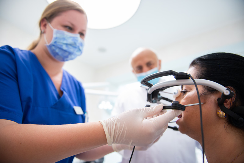
By means of a face-bow (fig.) the position of the maxilla to mandible (upper- to lower jaw), as well as their relation to the base of the scull, will be measured.
The results obtained by the employment of the face- bow will be used to simulate mandibular movements. By fitting plaster casts of the upper and lower jaw into an articulator, the masticatory movements of the patient can be reproduced. (fig.)
In our surgery we also make use of the computerised bite registry system Arcus Digma (fig.), which records the trajectories of the jaw by means of ultrasound impulses.
The T-scan-method may also be employed in order to conduct a four-dimensional analysis of the dental occlusion.
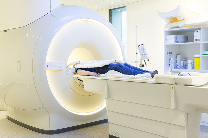
MRI
With the aid of MRI (magnetic resonance imaging) it is possible to depict tissue particularly rich in contrast. On suspicion of a TMD, the MRI can be employed as an imaging method in order to visualise the discs along with the osseous structure. A functional MRI can register the movements of the jaw and is therefore used to depict the position of the discs on opening and closing of the mouth.
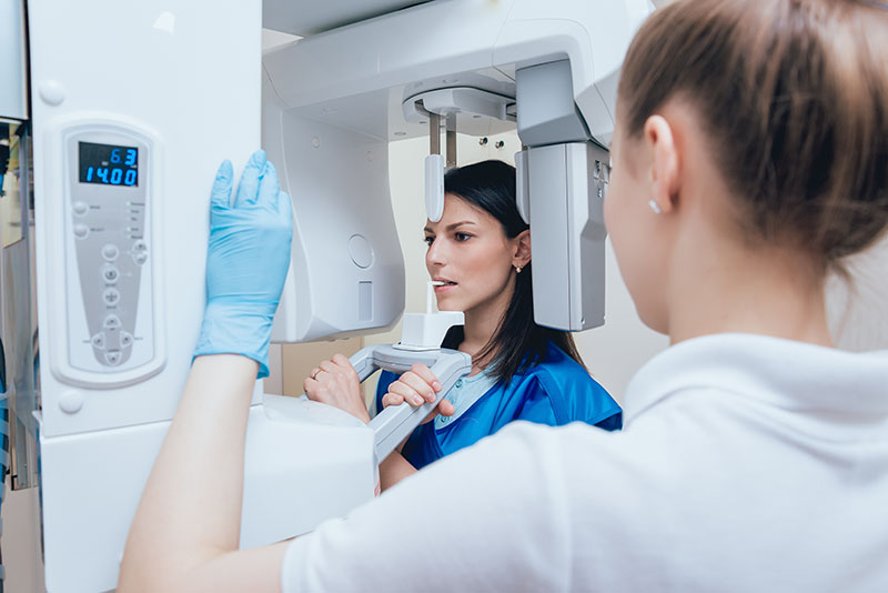
Digital Volume Tomography (DVT)
Unlike conventional two-dimensional X-ray methods, the digital volume tomography facilitates a three-dimensional depiction of even the most delicate anatomic structures. This modern X-ray method allows the compilation of high-resolution pictures of the osseous structure of the jaw. It can therefore be invaluable for the diagnosis of TMD. The DVT will be performed right at our dental surgery.
Therapy
We offer you the best treatment for a healthy and relaxed jaw!
The therapy process around TMD is as diverse as the diagnostics. A distinction is made here whether there is a physiological cause or not.
Splint therapy is sufficient in most cases as a treatment for TMD. There is also the possibility of electrical stimulation to relieve any muscular pain that may be present. Our interdisciplinary team of experts will take care of you throughout your treatment and across all departments.
An untreated TMD can worsen and produce increasingly severe symptoms. Our dentists and our interdisciplinary team of experts are ready to offer you comprehensive treatment in a timely manner, because an early treatment time can clearly have a positive effect on the course of therapy and save the sufferer a lot of pain.
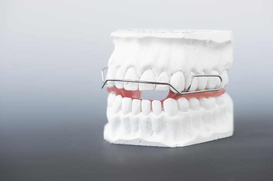
Dental Splint therapy
If a dental cause for the TMD has been diagnosed, a dental splint therapy (occlusal splint) will be applied. Generally, there is a differentiation between two different types of splints: functional and protective splints. Every clinical presentation calls for its own especially adjusted kind of therapeutic splint.
Functional splints are deployed as an individual therapeutic bite splint in order to bring and hold the temporomandibular joint condyles (fig.) in the desired therapeutic position.
Protective splints, such as mouth guards, are used to treat bruxism (unconscious teeth clenching) and usually serve to relieve the musculature of stress and protect the hard tooth tissue and/or the prosthetics (crowns, bridges etc.) from wear.
A mandibular advancement splint, a so called “snore guard”, can be applied in order to expand the throat during sleep. Therefore, snoring and slight cases of obstructive sleep apnoea can be successfully alleviated.
Electric Muscle Stimulation
It is possible to relieve the accompanying muscle pain of a TMD by application of electromedical stimulation current therapy. TENS (Transcutaneous Electrical Nerve Stimulation) works by stimulating nerves in the painful area by use of a specific electrical pulse, which can result in the relaxation of the affected musculature. This relaxation of the masticatory muscle effects pain relief in the affected regions. This treatment can be performed in our dental surgery with our own TENS device, which has been specifically adapted to aid in the treatment of TMD.
Interdisciplinary Team of Experts
If any abnormalities in relation to the clinical picture of TMD which are not directly related to dentistry should reveal themselves during our treatment of TMD, we will gladly refer you to an affiliated specialist. We are working closely together with highly qualified colleagues from the most diverse disciplines: e.g. neurology, neurosurgery, otorhinolaryngology, orthopaedics, internal medicine, radiology and ophthalmology.

Further Treatment Procedure
Usually, splints are sufficient for the treatment of TMD. But in some cases, the alignment of the maxillary and mandibular teeth has become incoherent in consequence of the alteration of the temporomandibular joints. This can be remedied by a correction of the occlusion.
Often, wearing a splint becomes dispensable after the new occlusion has consolidated itself. Concerning crowns, bridges, Table Tops, implants and veneers, our specifically trained prosthodontists (dentists) have the most advanced methods of treatment at their disposal.
